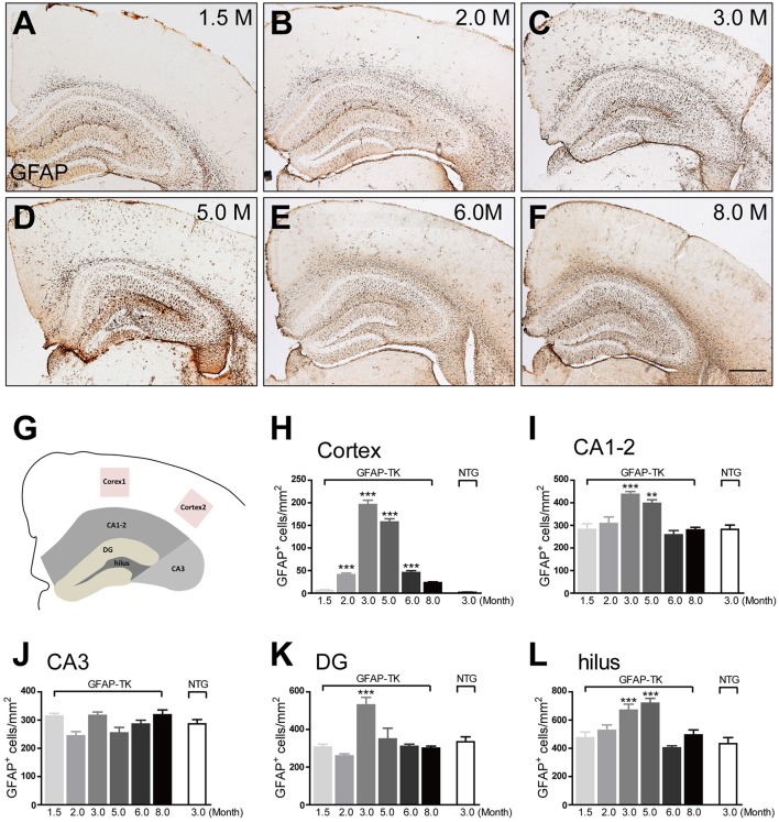
Figure 3.
The increased expression of GFAP in the cortex and hippocampus of 3-month-old TK-1 mice was found between 3 and 5 months old of age (A–F) Representative photomicrographs of GFAP+ cells in the cortex and hippocampus of 1.5-, 2-, 3-, 5-, 6-, and 8-month-old TK-1 mice. Scale bar, 500 μm. (G) Schematic depicting regions of the cortex and hippocampus analyzed for this study. (H) Quantification of the number of GFAP+ cells in the cortex of 1.5-, 2-, 3-, 5-, 6-, and 8-month-old TK-1 mice (n = 3 mice per group, 3 brain slices per mouse). (I–L) Quantification of the number of GFAP+ cells in the hippocampus of 1.5-, 2-, 3-, 5-, 6-, and 8-month-old TK-1 mice (n = 3 mice per group, 3 brain slices for each mouse). (I), CA1-2; (J), CA3; (K), DG; (L), hilus. Data represent mean ± SEM,*P < 0.05, **P < 0.01, ***P < 0.001 (one-way ANOVA with post hoc Turkey's multiple comparison test).

