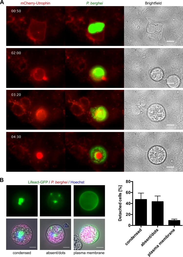
FIG 1 .
Host cell actin-plasma membrane linkage is lost during Plasmodium egress from host hepatocytes. (A) HeLa cells were transfected with a construct encoding mCherry-utrophin (red) and infected with GFP-expressing parasites (green). Shown are stills of a live-cell imaging movie that was started at 62 hpi. (B) Representative images of the different types of actin localization (condensed, absent or dots, and plasma membrane associated) in detached cells derived from primary Lifeact hepatocytes. Primary hepatocytes were isolated from a Lifeact mouse and infected with mCherry-expressing parasites (red). At 65 hpi, detached cells were harvested and analyzed by live-cell microscopy. Nuclei were visualized with Hoechst 33342 (blue). The actin cytoskeleton is shown in green. The relative percentage of each type of actin localization is shown as the mean and the standard error of the mean on the right and was quantified in three independent experiments in which a total of 189 detached cells were analyzed. All scale bars, 10 µm. See also Movies S1 and S2.

