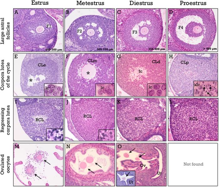
Figure 5. Hallmarks of the mouse estrous cycle.
These include large antral follicles (A–D), corpora lutea of the current cycle (E–H), one cycle-old (I–L) regressing corpora lutea, and presence of ovulated oocytes in the oviduct (M–O). Corpora lutea of the current cycle show non-luteinized granulosa cells (lc) in estrus (CLe in panel E), non-fully luteinized cells in metestrus (CLm in panel F), with a prominent vascular pattern and mitotic figures, and full luteinization in diestrus (CLd in panel G). An empty cavity (denoted by asterisks) is frequently observed in estrus and metestrus, whereas a fibrous center (fc in G) is frequent in diestrus. The presence of apoptotic cells is characteristic of the CL in proestrus (arrows in panel H). Regressing corpora lutea (RCL in panels I–L) show an increasing ratio of stromal to steroidogenic cells, progressively decreasing size and are practically demised at the end of the cycle. A specific signal for estrus is the presence of cumulus-oocyte-complexes (arrows in panel M) at the ampulla of the oviduct. Nude oocytes are located at the isthmus in metestrus (arrows in panel N), at the utero-tubal junction (arrows in panel O) or at the tip of the uterine horn (arrow in the inset in O) in diestrus, and are not found in proestrus. Hematoxylin and eosin staining.

