Fig. 5.

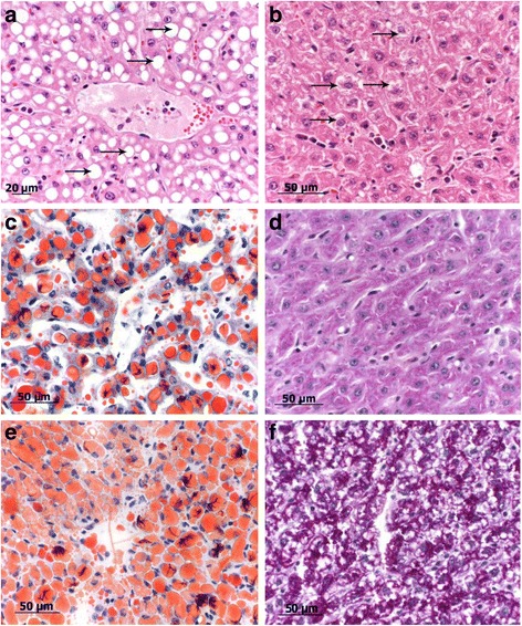
Bright field images, comparison different morphology of fatty vacuoles and glycogen depositions of formalin fixed and snap frozen liver samples, H&E, PAS and ORO staining. a large, clear demarcated fatty vacuoles intracytoplasmatically in hepatocytes displacing the nucleus excentrically. b Intracytoplasmatical glycogen deposition (arrows) in swollen hepatocytes showing a foamy brightened cytoplasm. c ORO staining of formalin fixed tissue sample, showing clear red stained fatty vacuoles diffusely distributed in the liver lobule. d PAS staining of glycogen deposition of formalin fixed tissue samples. e ORO staining of snap frozen liver sample, diffuse red coloration of hepatocytes, unclear cell borders, sinusoids indistinct and difficult to localize. Distribution of fatty vacuoles similar to formalin fixed tissue. f PAS staining of snap frozen liver samples, diffuse pinkish coloration of hepatocytes, unclear cell borders, sinusoids indistinct and difficult to localize. Distribution of glycogen depositions similar to formalin fixed tissue
