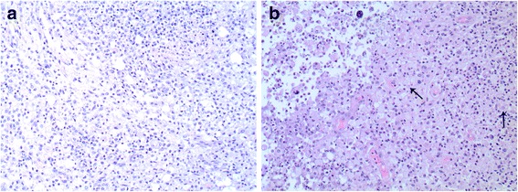
Fig. 2.

Xanthogranulomatous pyelonephritis and coexisting malakoplakia of the left kidney. Histopathological examination of the surgical specimen with hematoxylin and eosin staining reveals microscopic features of chronic inflammation. a Heavy lymphoplasmacytic infiltration. b Inflammatory infiltrates with layered sheets of histiocytes. Michaelis–Gutmann bodies pathognomonic for malakoplakia (granulomatous inflammation of the genitourinary tract) are indicated by arrows
