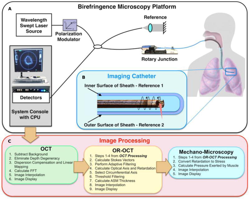
Fig 1. Birefringence Microscopy Platform schematic.

(A) A schematic of the Birefringence Microscopy Platform. The fiber-based imaging system utilizes a wavelength swept-source. (B) Depiction of the endoscopic imaging catheter. A ball lens was made from a fiber tip and was angle polished for side-viewing. The imaging pitch is determined by the ratio of the rotation rate to pullback speed, and a dual-layer torque coil protects the imaging fiber and allows for rotation with minimal distortion. The inner and outer surfaces of the birefringent polymer sheath are used to obtain the reference orientation for in vivo OR-OCT imaging. (C) Outline of the processing steps used for obtaining OCT, OR-OCT, and Mechano-Microscopy images. A complete description of the components and processing can be found in the Supplementary Materials.
