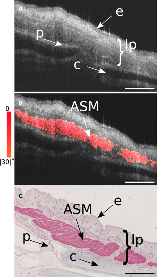
Fig. 2. Segment of pig trachea where smooth muscle connects two cartilage plates.

(A) Structural OCT image of a segment of swine trachea that includes trachealis muscle. This segment of tissue was bisected and removed from the remaining trachea and was imaged using a bench-top scanner with the circumferential axis oriented in line with the primary image-scanning axis (see Fig. S7 - inset for bisection schematic). Features such as the epithelium, lamina propria, perichondrium, and cartilage rings are well defined, but the smooth muscle is often indistinguishable from surrounding glandular and connective tissue. (B) Using our birefringence microscopy platform we are able to separate out the circumferentially oriented (+/− 30°) birefringent tissue that corresponds to the smooth muscle. (C) The OCT image was matched to histology stained with alpha smooth muscle actin (αSMA). Comparison between the OR-OCT image and the αSMA stained histology reveals the high degree of isolation and sensitivity our platform is capable of for identifying smooth muscle. lp, lamina propria; e, epithelium; p, perichondrium; c, cartilage. Scale bars, 500 μm.
