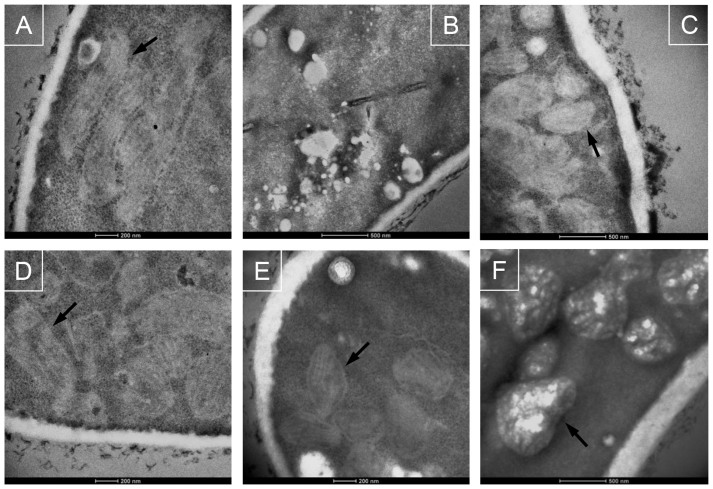
Figure 2. Ultrastructural morphology of A. fumigatus hyphae in the presence of phenazines.
(A) A. fumigatus conidia incubated for 20 h growth at 30°C in the presence of 1% DMSO. (B–F) A. fumigatus conidia incubated for 18 h growth at 37°C in the presence of 1 mM PYO (B, C), 125 μM PCN (D), 62.5 μM 1-HP (E) or 2 mM PCA (F). Arrows show the mitochondria in A. fumigatus cells.

