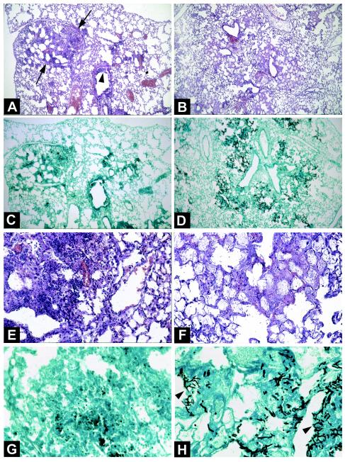
FIG. 5.
Lung histological features of corticosteroid- and chemotherapy-treated mice infected with A. fumigatus. (A, C, E, and G) Lung sections from a corticosteroid-treated mouse 72 h after intratracheal inoculation with 107 conidia. (A) Hematoxylin-eosin stain; magnification, ×60. (E) Hematoxylin-eosin stain; magnification, ×250. A large focus of pneumonia (A, arrows) and exudative bronchiolitis (A, arrowhead) with the destruction of bronchi and alveolae can be seen. Hemorrhagic necrosis and numerous inflammatory cells, especially PMN, also can be seen (E). (C) Methenamine silver stain; magnification, ×60. (G) Methenamine silver stain; magnification, ×400. A few conidia and very few hyphae within the lesions of necrosis can be seen. (B, D, F, and H) Lung sections from a chemotherapy-treated mouse 48 h after intratracheal inoculation with 107 conidia. (B) Hematoxylin-eosin stain; magnification, ×60. (F) Hematoxylin-eosin stain; magnification, ×250. Lesions of bronchiolitis and diffuse pneumonia with edema and congestion within alveolae (B) but without an inflammatory exudate of any cell type, including PMN, can be seen. (D) Methenamine silver stain; magnification, ×60. (H) Methenamine silver stain; magnification, ×400. Several foci of bronchocentric necrosis containing numerous hyphae (H, arrowheads) can be seen. Invasion of alveolae by numerous hyphae of A. fumigatus also can be seen. Note the absence of an inflammatory exudate.

