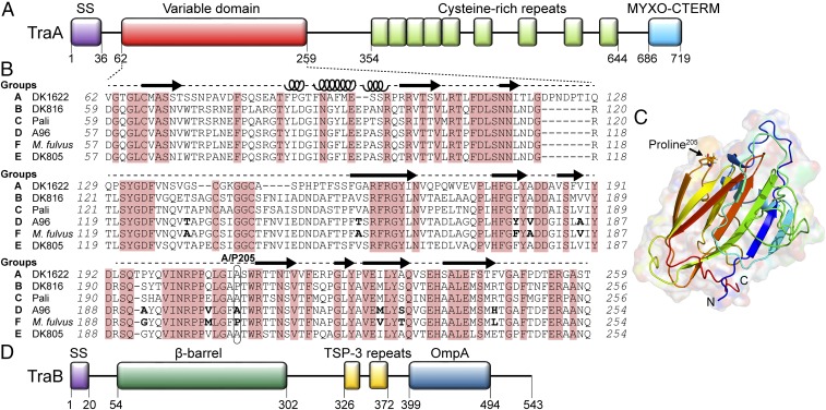
Fig. 1.
TraA contains a variable domain that determines recognition specificity. (A) Domain architecture of TraA: Type I signal sequence (SS); Cys-rich region and MYXO-CTERM are a putative stalk and a sorting tag, respectively (11). (B) Sequence alignment of the TraA variable domain from six recognition groups. Conserved residues are shaded red. Amino acid differences between the TraAA96 and TraAM. fulvus are in bold. Predicted secondary structures by I-TASSER (22) are indicated (loops, α-helices; arrows, β-strands). (C) Modeled 3D structure of the TraAM. fulvus variable domain by I-TASSER where the highest C-score was selected. (D) Domain architecture of TraB; Thrombospondin type 3 (TSP-3, Pfam02412). β-Barrel prediction was made with BOCTOPUS2 (32).

