Fig. 4.
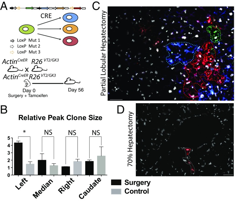
The neonatal mouse liver regenerates through clonal hepatocyte proliferation. (A) Schematic of the Cre-dependent “Rainbow” reporter and experimental design: ActinCreERT2R26 VT2/GK3 pups underwent partial lobular hepatectomy at day 0 and were treated with tamoxifen. Mice were followed for 56 d. (B) Analysis of relative peak clone size per lobe in mice 56 d postsurgery compared with age-matched controls. Values are means ± SEM; *P = 0.05, **P < 0.005, ***P < 0.0005, n = 1,445. Clones ranged from single cells to 408 (max). (C) Representative image across three channels (GFP, RFP, and CFP) showing large multicolor clones in a 56-d postpartial hepatectomy day 0.5 mouse merged with Hoechst 33342. (D) Representative image showing smaller clones in an adult undergoing classical partial hepatectomy merged with Hoechst 33342. (Scale bars, 100 μm.)

