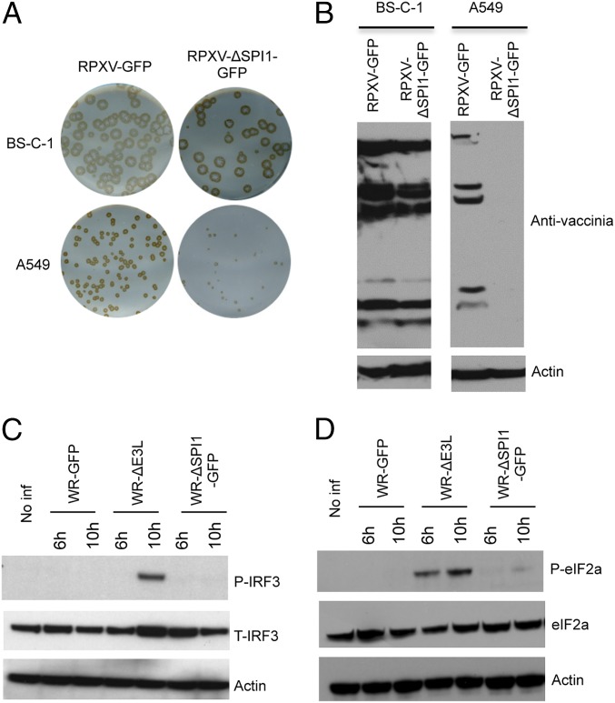
Fig. 1.
Host-range restriction of SPI-1 mutants. (A) Plaque formation. BS-C-1 and A549 cells were infected with control RPXV-GFP or SPI-1 deletion mutant RPXV-ΔSPI1-GFP. Plaques formed in 72 h were detected by immunostaining with rabbit polyclonal anti-VACV antibody. (B) Immunoblots of viral proteins. Proteins from BS-C-1 and A549 cells infected for 28 h with RPXV-GFP or RPXV-ΔSPI1-GFP were resolved by polyacrylamide gel electrophoresis, transferred to a membrane, and probed with polyclonal antibody to VACV and actin as a loading control. (C) Immunoblot of IRF3. Proteins from A549 cells that were noninfected (No Inf) or infected with wild-type VACV strain WR (WR), a VACV E3 deletion mutant (ΔE3L), or VACV-ΔSPI1-GFP (ΔSPI1-GFP) for 6 or 10 h, as indicated, were analyzed as in B and probed with antibody to phosphorylated IRF3 (P-IRF3), total IRF3 (T-IRF3), or actin. (D) Immunoblot of eIF2α. Same as C except that blots were probed with antibody to phosphorylated eIF2α (P-eIF2α) or total eIF2α protein (eIF2α).

