Fig. 2.
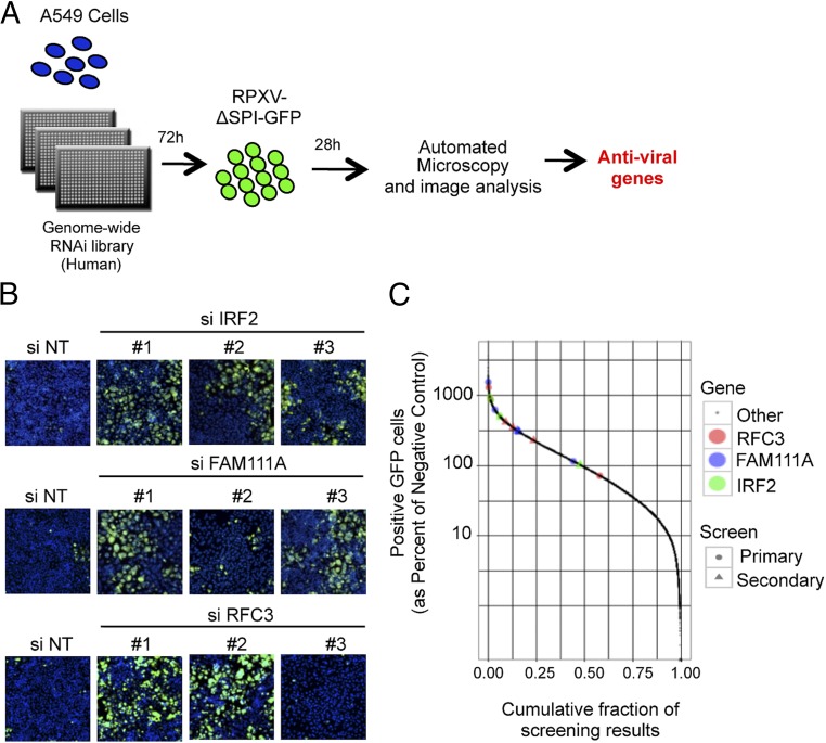
Genome-wide siRNA screen. (A) Schematic of the human genome-wide screen. A549 cells in a 384-well plate were reverse transfected for 72 h with the Silencer Select siRNA library from Ambion, infected with 0.01 PFU per cell of RPXV-ΔSPI1-GFP for 28 h, fixed, and screened for cells that stained with Hoechst and exhibited GFP fluorescence. Antiviral genes were determined by increased number of cells with GFP fluorescence compared with median. (B) Images of the siRNA-transfected and virus-infected cells from the primary screen. NT stands for nontargeting siRNA. Hoechst stain, blue; GFP, green. (Magnification: 10×.) (C) The percentages of fluorescent cells from the primary and secondary screens for individual IRF2, FAM111A, and RFC3 siRNAs (divided by the percentages of fluorescent cells for negative controls) compared with siRNAs for all other genes. Color and symbol keys for siRNAs on right.

