Fig. 5.
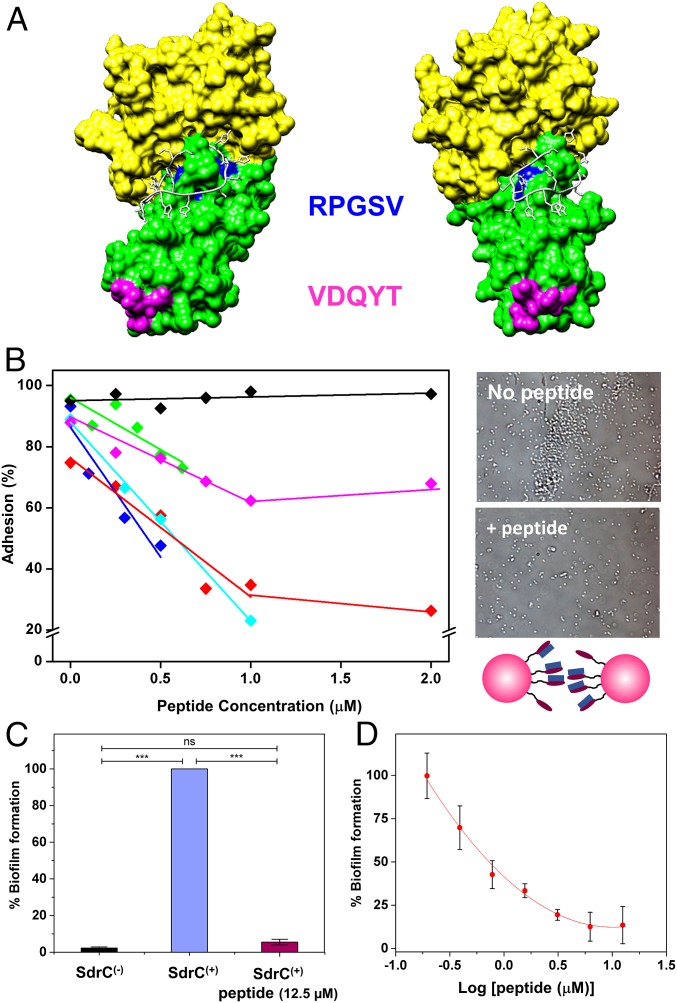
Blocking cell–cell adhesion forces using β-neurexin–derived peptide. (A) Molecular model of the SdrC–peptide interaction. SdrC N2 and N3 subdomains are colored green and yellow, respectively. The RPGSV and VDQYT sequences are colored blue and pink, respectively. The peptide is shown in white in stick format. The image shown (Right) is rotated 42° compared with the view (Left). (B) Inhibition of cell–cell adhesion forces. Variation of the adhesion probability measured by SCFS for five L. lactis SdrC(+) cell pairs (different colors) upon addition of β-neurexin–derived peptide at increasing concentrations. As a control, a scrambled peptide was tested (black symbols). (B, Right) Optical images showing L. lactis SdrC(+) bacteria before and after addition of 12.5 µM peptide. (C and D) Inhibition of SdrC-mediated biofilm formation. SdrC(+) and SdrC(−) cells were allowed to form a biofilm at 30 °C for 24 h and stained with crystal violet, and the absorbance was measured at 570 nm. Values are expressed as the percentage of the value for wells containing SdrC(+) bacteria without peptide. Results shown are the mean values of triplicate samples, and error bars represent the SEM; P values were calculated using Student's t test. ***P < 0.001; ns, not significant. In D, varying concentrations of the peptide (0.195 to 12.5 µM) were added to the diluted bacteria before addition to the microtiter plate.

