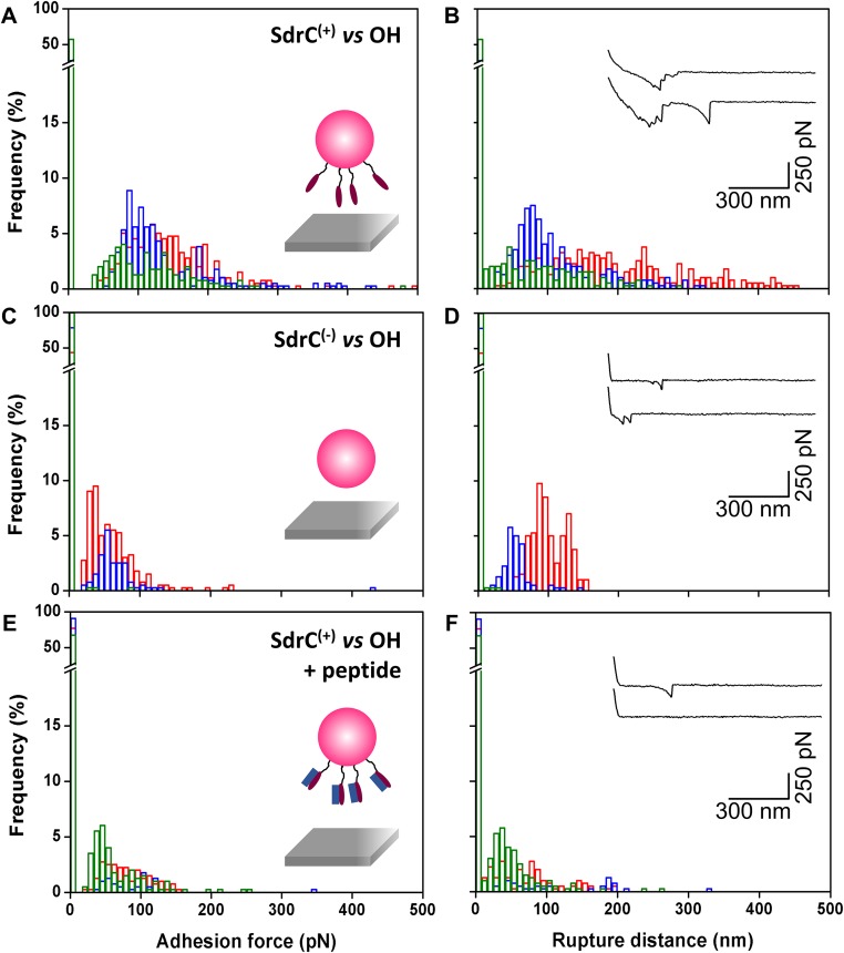
Fig. S4.
Forces guiding bacterial attachment to hydrophilic surfaces. (A–D) Adhesion force (A and C) and rupture distance (B and D) histograms obtained in PBS between three different L. lactis SdrC(+) cells (A and B) or L. lactis SdrC(−) cells (C and D) and hydrophilic, hydroxyl-terminated substrates. (E and F) Force data were collected for L. lactis SdrC(+) cells in the presence of β-neurexin–derived peptide (2 µM). Curves were obtained using a contact time of 100 ms, applied force of 250 pN, and approach and retraction speed of 1.0 µm/s.

