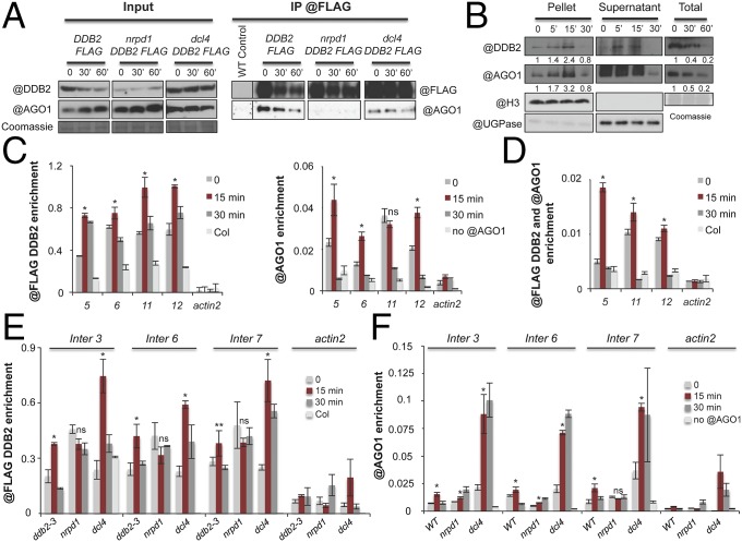
Fig. 5.
DDB2 AGO1 homeostasis and DDB2−AGO1 complex. (A) In vivo pull-down of AGO1 with DDB2-FLAG protein upon UV-C exposure; ddb2-2/DDB2-FLAG, nrpd1 ddb2-2/DDB2-FLAG, and dcl4 ddb2-2/DDB2-FLAG expressing plants were used for IP assays using anti-FLAG antibody. WT (No) plants were used as negative control. Coomassie blue staining of the blot is shown. (B) Immunoblot analysis of DDB2 and AGO1 protein contents upon UV-C exposure in chromatin (pellet), supernatant, and total extracts from WT plants. Anti-histone H3 and anti-UGPase antibodies were used as controls for insoluble (Pellet; chromatin) and soluble fractions (Supernatant), respectively. Signal intensity relative to H3 or Coomassie is indicated below each lane. Coomassie blue staining of the blot is shown. (C) ChIP of (Left) DDB2-FLAG and (Right) AGO1, upon UV-C exposure, at four hot spots in ddb2-3/DDB2-FLAG expressing plants using anti-FLAG and anti-AGO1 antibodies, respectively. As negative control for DDB2 ChIP, WT (Col) plants were used with anti-FLAG antibody as well as actin2 region. As negative control for AGO1 ChIP, WT (Col) plants were used with protein A magnetic beads as well as actin2 region. Data are presented as enrichment (±SD) of the IP signal and are representative of three independent biological replicates; t test *P < 0.01; ns, nonsignificant compared with time point 0. (D) Tandem ChIP (Tandem-ChIP) of DDB2-FLAG and AGO1, upon UV-C exposure, at three hot spots in ddb2-3/DDB2-FLAG expressing plants using anti-FLAG antibody followed by anti-AGO1 antibody. As negative control for ChIP, WT (Col) plants were used as well as actin2 region. Data are presented as enrichment (±SD) of the IP signal and are representative of two independent biological replicates; t test *P < 0.01 compared with time point 0. (E) ChIP of DDB2-FLAG upon UV-C exposure, at three hot spots in ddb2-3 DDB2-FLAG, nrdp1 DDB2-FLAG, and dcl4 DDB2-FLAG expressing plants using anti-FLAG antibody. As negative control for DDB2 ChIP, WT (Col) plants were used with anti-FLAG antibody as well as actin2 region. Data are presented as enrichment (±SD) of the IP signal and are representative of three independent biological replicates; t test *P < 0.01; **P < 0.05; ns, nonsignificant compared with time point 0. (F) ChIP of AGO1, upon UV-C exposure, at three hot spots in WT, dcl4, and nrpd1 plants using anti-AGO1 antibody. As negative control for AGO1 ChIP, WT (Col) plants were used with protein A magnetic beads as well as actin2 region. Data are presented as enrichment (±SD) of the IP signal and are representative of three independent biological replicates; t test *P < 0.01; ns, nonsignificant compared with time point 0.

