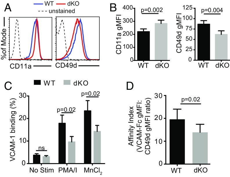
Fig. 4.
Activated EVL/VASP dKO CD4 T cells have a deficit in α4 integrin (CD49d) expression and function. (A) Examples of CD11a and CD49d expression on WT and EVL/VASP dKO activated T cells by flow cytometry. (B) Quantification of CD11a and CD49d surface expression in activated WT and dKO T cells; data shown as gMFI. (C) CD49d function measured as soluble VCAM-1 binding to T cells in response to the indicated stimuli (MnCl2: manganese chloride; PMA/I: PMA and ionomycin). (D) Affinity for VCAM-1 calculated as PMA/ionomycin-elicited VCAM-1 binding normalized to surface expression of CD49d by gMFI. Data in A are representative of 10 independent experiments; data in B are the average of ten experiments; data in C and D are the average of three independent experiments. Error bars are SEM. All P values are paired t tests. ns, not significant.

