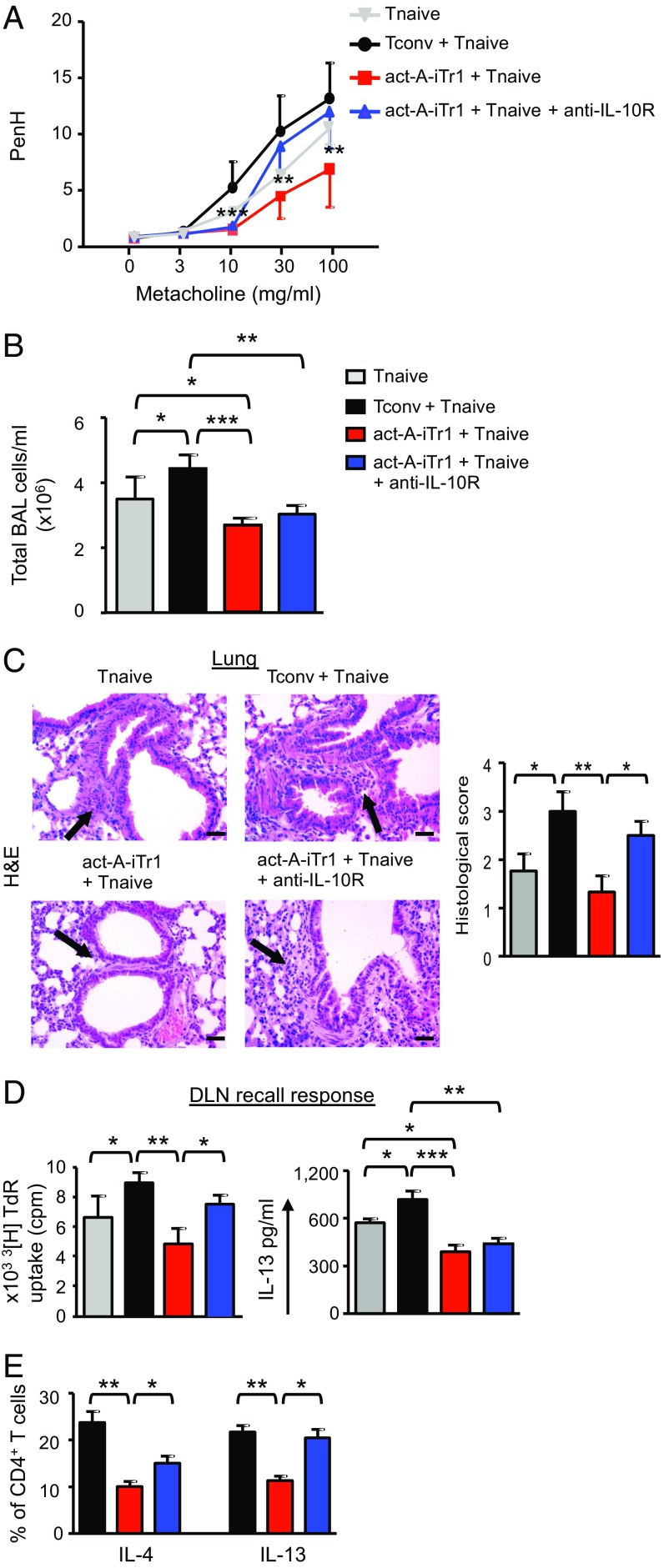
Fig. 6.
In vivo administration of human act-A–iTr1 cells prevents allergic airway disease. (A) AHR is depicted. Enhanced pause (PenH) results are expressed as mean ± SEM. Data were analyzed by two-way ANOVA for repeated measures, followed by the Student’s t test, and are pooled from two to three independent experiments (n = 4–6 mice per group). (B) Total BAL cell numbers are shown. (C) Representative photomicrographs and histological scores of H&E-stained lung sections. (Scale bars: 50 μm). (D) DLN cells were harvested at the time of euthanasia and restimulated with P. pratense. 3[H]Thymidine incorporation and IL-13 release in culture supernatants are presented. Data are mean ± SEM of triplicate wells. (E) Percentages of cytokine-producing human CD4+ T cells in the DLNs are shown. Data are representative of two to three independent experiments (n = 4–6 mice per group). Statistical analysis was performed by the Student’s t test (*P < 0.05; **P < 0.01; ***P < 0.001).

