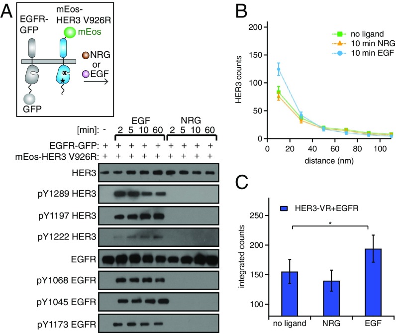
Fig. 5.
In the EGF-induced clusters, HER3 is phosphorylated by EGFR independently of asymmetric kinase dimerization. (A, Upper) Cartoon depicting the EGFR–GFP and mEos–HER3–V926R receptors. (Lower) Western blot analysis of the lysates from NR6 cells stably expressing mEos–HER3–V926R and EGFR–GFP. The lysates were collected after stimulation with 10 nM EGF or 10 nM NRG for the indicated periods of time. (B and C) Pairwise distance histograms (B) and integrated counts from those histograms calculated from the STORM images (C) (as described in Fig. 2) for cells coexpressing the mutant mEos–HER3–V926R and EGFR–GFP and stimulated with 10 nM EGF or 10 nM NRG for 10 min. *P < 0.05.

