
Figure 4.
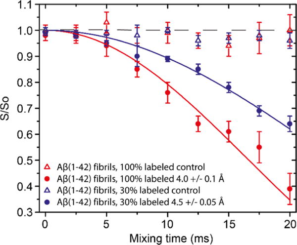
FS-REDOR of AβM01–42 fibrils recorded at 750 MHz, T = 277 K, and ωr/2π = 8 kHz with ω1H/2π = 83 kHz 1H decoupling field applied during acquisition. The Gaussian selective π pulse on 13C was 0.6 ms long and set on the resonance of A42−13COO−. For 15N, ω1S/2π = 33 kHz during REDOR and set to the resonance of the Nζ of K28. The S and S0 signals were measured with and without the 15N selective pulse, respectively. The curve fits show that a salt bridge exists between K28 and A42 with a distance of 4.0 Å in the 100% labeled sample and 4.5 Å in the 30% sample. Intermolecular contacts between the PIR fibrils are responsible for the discrepancy in the dephasing observed.