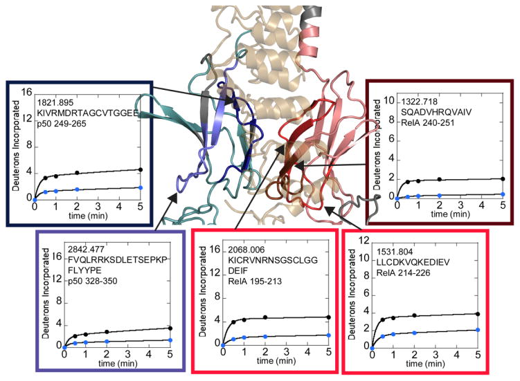
Figure 3.
HDXMS analysis of free NFκB (black circles) compared to IκBα-bound NFκB (blue circles). The interface regions within the dimerization domain subdomains that are protected upon IκBα binding are shown on a model of the NFκB-IκBα structure. RelA is shown in salmon with protected regions in hues of red. Only the dark red region is directly contacting IκBα. P50 is shown in teal with protected regions in hues of blue. Only the dark blue region is directly contacting the IκBα. Regions of each protein that were not covered in the HDXMS analysis are grey. The deuterium uptake plots are positioned near and arrows point to the corresponding structural region. The plots are boxed with colors similar to those used to denote direct and indirect protection on the structure.

