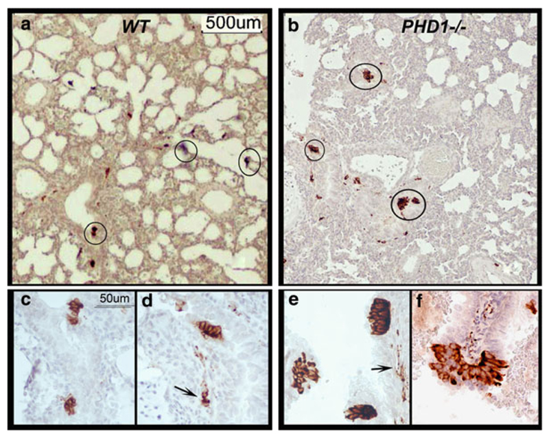
Fig. 21.1.
NEB in lungs of WT control and PHD-1 deficient mice visualized using immunoperoxidase method with anti-SV2 antibody. (a) Low magnification view of lung section from neonatal (P2) WT mice showing positive NEB in small airways (circled). (b) Section of lung from neonatal ( P2 ) PHD-1 deficient mouse lung at the same magnification as in (a) showing more prominent NEBs (circled) in similar distribution. (c) Close up of two NEBs consisting of small clusters of immunopositive cells in epithelium of small airway in lung from WT mouse. (d) Another small NEB in the same sample as in (c) with immunopositive nerve fibers (arrow) in submucosa. (e) Higher magnification view from sample (b) with three prominent NEBs forming compact intraepithelial corpuscles and immunoreactive nerve fibres in submucosa (arrow). (f) Close up of large, hyperplastic NEB from sample (b) situated at airway bifurcation and protruding into airway lumen

