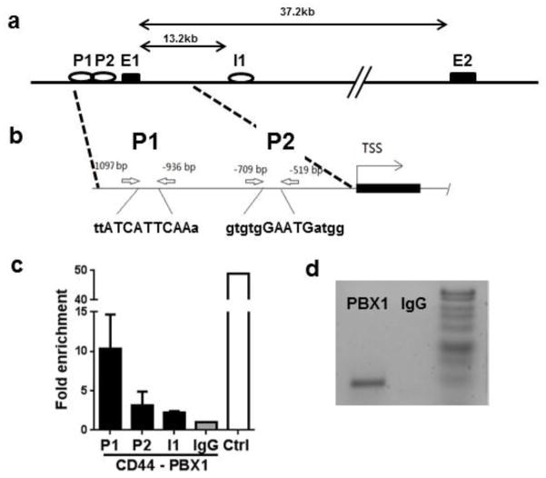
Fig. 1. PBX1 binds multiple sites in the CD44 gene.
(a) Map of the CD44 gene with the location of the putative PBX1 binding sites in intron 1 (I1) and the promoter (P). (b) Details of two putative promoter PBX1 binding sites (P1 and P2) as well as their corresponding PBX1 binding sequences in upper case. (c) Chromatin from Jurkat T cells was immunoprecipated with antibody against PBX1 or IgG control, and amplified by qPCR with primers flanking each of the 2 promoter sites and the intron 1 site. A positive control (Ctrl) was provided by immunoprecipitation with a POL2 antibody and amplification of the POL2 binding site in the GAPDH promoter. CHiP-qPCR results are expressed in fold enrichment relative to the IgG negative control (N = 3). (d) Representative CHiP-PCR product at the P1 site.

