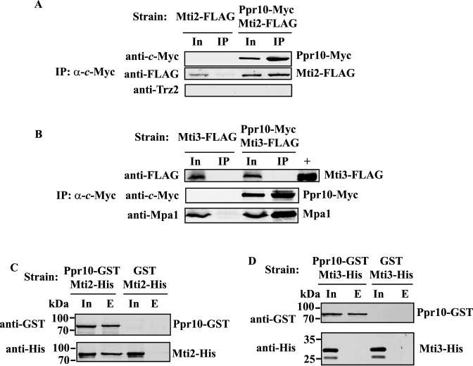
Figure 10.
Ppr10 interacts with Mti2 but not with Mti3 in vivo and in vitro. (A and B) Ppr10 co-immunoprecipitates with Mti2, but not with Mti3. Extracts of cells expressing chromosomally encoded (A) Ppr10-Myc and Mti2-FLAG or (B) Ppr10-Myc and Mti3-FLAG were used for IP with anti-c-Myc agarose beads. Control IPs were also performed on extracts of cells expressing chromosomally encoded (A) Mti2-FLAG or (B) Mti3-FLAG. Extracts were prepared by glass bead disruption. IPs were performed in the same way as described in Figure 5, except that the beads were washed under moderate stringent conditions to prevent dissociation of weakly bound proteins (see Materials and Methods for details). Extracts and IPs were subjected to western blotting with indicated Abs. Input (In) lanes contain 4% of the extracts used for IP. Lane + contains affinity purified Mti3-FLAG served as the positive control. Anti-Mpa1 Ab was used as a positive control. (C and D) Ppr10 pulls down Mti2, but not Mti3. Whole cell lysates of E. coli cells co-expressing GST or Ppr10-GST with (C) Mti2-His, or (D) Mti3-His were incubated with glutathione resin, and proteins bound to GST were analyzed by western blotting with anti-GST and anti-His Abs. In, Input (1% of total protein), E, bound proteins.

