Figure 5.
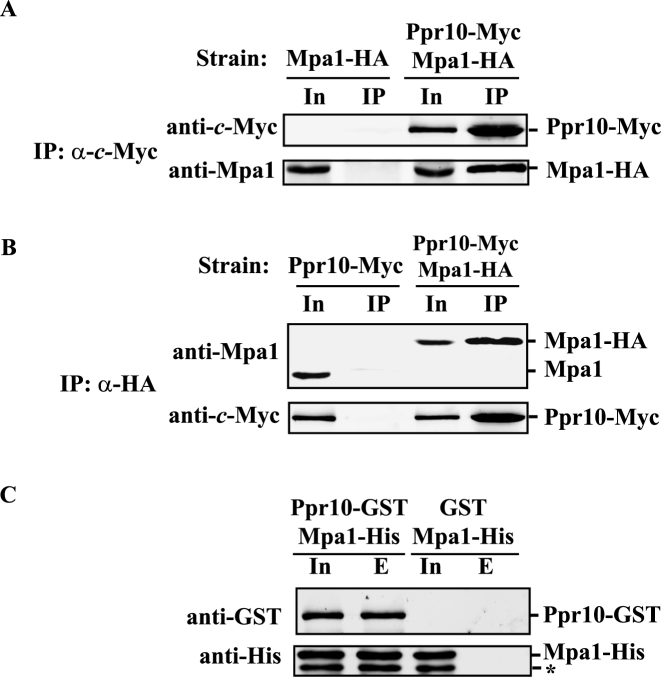
Ppr10 interacts with Mpa1 both in vivo and in vitro. (A) Co-immunoprecipitation (co-IP) of Ppr10 with Mpa1. Cells expressing chromosomally encoded Ppr10-Myc and Mpa1-HA were grown to mid-log phase in YES medium, lysed by glass bead beating and subjected to anti-c-Myc IPs. Extracts and IP were analyzed by western blotting with indicated Abs. 4% of the input extract (In) was run to show the expression level of each protein. An extract from WT cells expressing untagged Ppr10 and chromosomally encoded Mpa1-HA was used as a control. (B) Reciprocal co-IP of Ppr10 and Mpa1. The same extract in (A) was subjected to anti-HA IPs. Extracts and immunoprecipitates were analyzed by western blotting with indicated Abs. The amount of input (In) is 4% of the lysate used for IP. As a control, IP was performed on an extract from WT cells expressing untagged Mpa1 and chromosomally encoded Ppr10-Myc. (C) Ppr10 directly interacts with Mpa1. E. coli extracts expressing Ppr10-GST and Mpa1-His or GST and Mpa1-His were incubated with glutathione resin. Input (In, 3% of total protein) and proteins bound to GST (lane E) were analyzed by western blotting. The asterisk depicts a degradation product from Mpa1.

