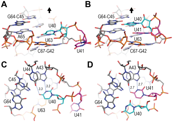
Figure 3.
The uridines at the 7SK motif. The (GAUC) repeat and uridines U40 (cyan), U41 (magenta) and U63 (orange). (A) Axial view of conformation OUT in molecule A (Os-map). The black arrow represents the helical axis and shows the direction of the apical loop. (B) Same view for conformation IN. (C) Perpendicular view for conformation OUT. (D) Perpendicular view for conformation IN. H-bonds of the Hoogsteen base-pair are indicated with dotted lines.

