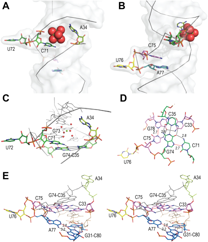
Figure 4.
The central region. (A) Global view of the central region (from native-map, molecule B), with single-stranded nucleotides A34 (anis), C71 (green), U72 (dark green), C75 (pink), U76 (yellow) and A77 (blue) in stick, showing the magnesium site (Mg, green sphere) surrounded by water molecules (red spheres). (B) Same view turned 90° about the helical axis. (C) Details of C71 interaction with base-pair G74-C35 (green). Dotted lines indicate H-bond, in red if protonation is required. (D) Top view of the triples on either sides of the CUA internal loop involving C71, with G74-C35 (green) and C75, with G78-C33 (pink). (E) Stereo view of the CUA internal loop showing C75 interaction with base-pair G78-C33 (pink) and A77 interaction with base-pair G31-C80 (blue). The intervening base-pair in thin lines is U32oG79.

