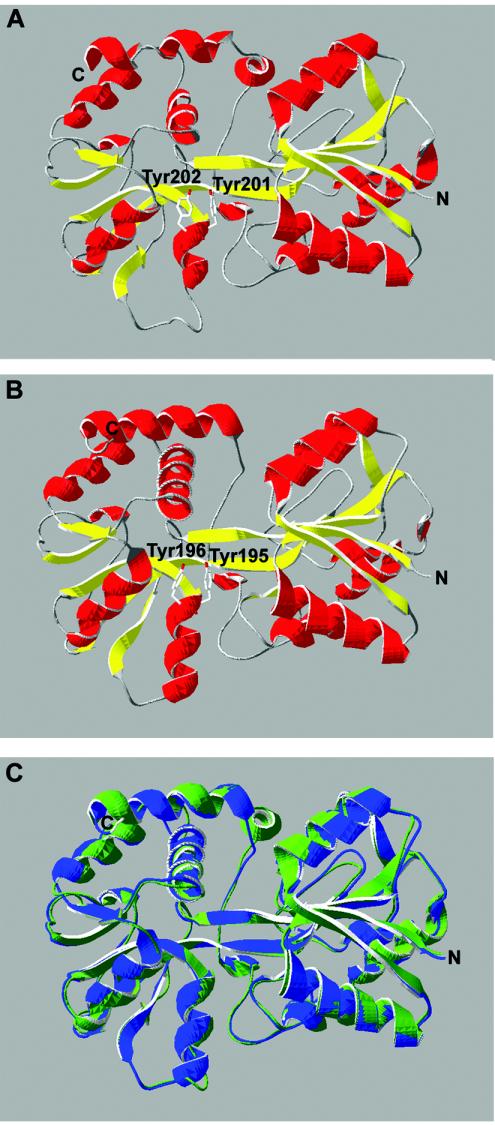
FIG. 3.
Three-dimensional structural modeling of E. canis Fbp using SWISS-MODEL. The putative structure of E. canis Fbp (A) is compared to the crystal structure of N. gonorrhoeae Fbp (B). Colors correspond to secondary structure: red, α-helix; yellow, β-pleated sheet. (C) Structural overlay of E. canis Fbp (green) with the crystal structure of N. gonorrhoeae Fbp (blue).

