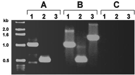
FIG. 5.
RT-PCR of E. canis fbp (lanes 1), E. canis membrane permease (lanes 2), and the intergenic region spanning the area from the 3′ end of fbp to the 5′ start of membrane permease (lanes 3) with either RNA from E. canis-infected DH82 cells (A), E. canis genomic DNA (B), or a control without RT (C).

