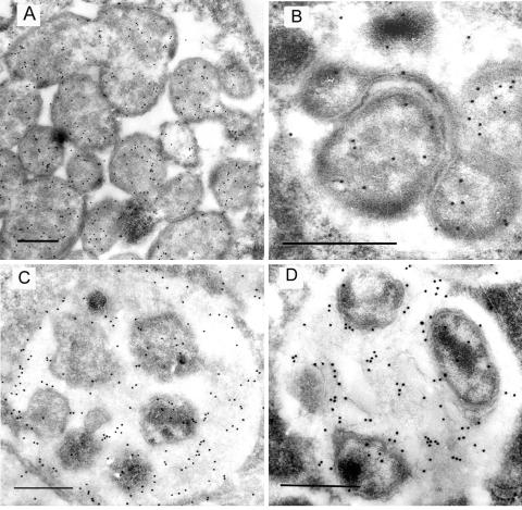
FIG. 7.
Immunogold-labeled electron micrographs of E. canis and E. chaffeensis with anti-E. canis recombinant Fbp. (A and B) Localization of Fbp in the E. canis (A) and E. chaffeensis (B) reticulate morphological form. (C and D) Localization of Fbp in the E. canis (C) and E. chaffeensis (D) dense-cored morphological form. Bars, 0.5 μm.

