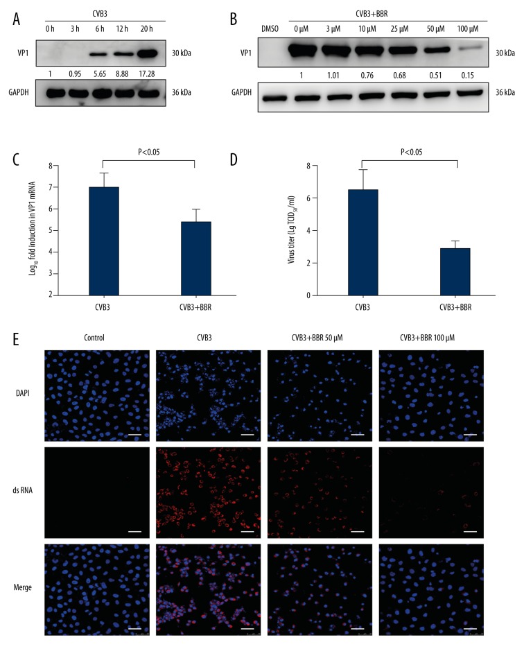
Figure 2.
BBR inhibits CVB3 replication in vitro. (A) HeLa cells were infected with CVB3 at 3 MOI for 1 h. Cell lysates were collected at the indicated times (3 h, 6 h, 12 h, 20 h) post-infection. Western blot analysis was performed to detect the expression of virus VP1 protein. (B) At 1 h after HeLa cells were infected with CVB3 (MOI=3), they were treated with BBR at the indicated concentrations (3 μM, 10 μM, 25 μM, 50 μM, 100 μM) for 20 h. Cell lysates were harvested and subjected to further analyses. Western blot analysis was performed to evaluate the expression of virus VP1 protein. (C) At 1 h after HeLa cells were infected with CVB3 (MOI=3), they were treated with BBR (100 μM) for 20 h. Real-time PCR analysis was performed to evaluate the levels of virus vp1 mRNA. (D) The viral titers after BBR (100 μM) treatment were determined by the Reed-Muench method. (E) IF staining was performed to detect the production of virus dsRNA during virus replication (dsRNA, red; nuclei, blue). The scale bar is 25 μm. Representative data from triplicate experiments are shown. Data show mean ±SD from 3 independent experiments. GAPDH was used as a protein loading control or endogenous reference gene.

