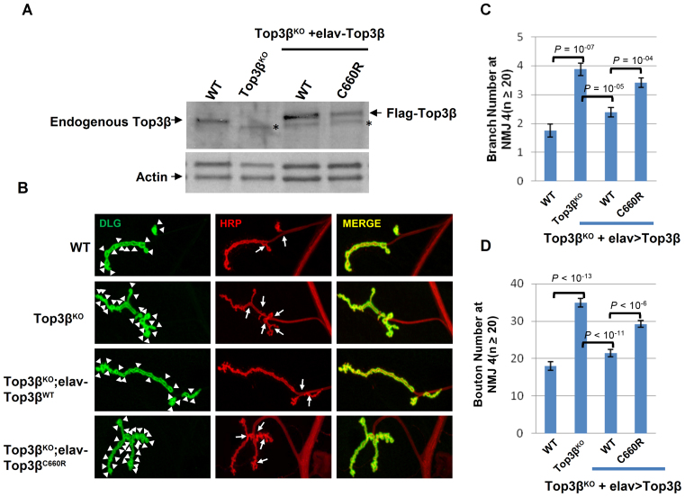
Figure 4.
A de novo SNV of Top3β from an autism patient is defective in promoting synapse formation in Drosophila. (A) Western blot images show expression levels of Top3β wild type and mutant proteins in extracts of adult brains from the indicated genotypes. (B) Representative immunofluorescence images of NMJs at muscle 4 (NMJ4) of wandering third instar Drosophila larvae of different genotypes as indicated. The NMJ4 was co-labeled with a presynaptic marker (anti-HRP, red) and a postsynaptic marker (anti-DLG, green). The arrowheads mark synaptic boutons, and the arrows mark branches. (C and D) Quantification of synaptic branches and boutons at NMJ4 from segments 3, 4 and 5 of both sides of wandering third instar larvae (n ≥ 20). The graphs show the means of the boutons and branches, and error bars represent standard errors of mean from three independent experiments. The P-values shown above each bar were calculated using Student's t-test.

