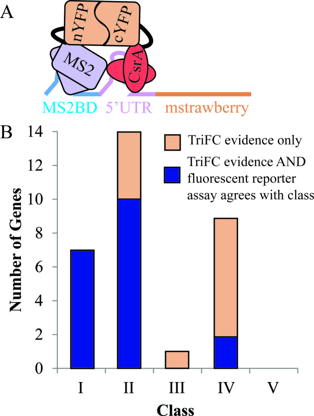
Figure 5.
TriFC data demonstrate interactions between CsrA and the 5΄ UTR of selected potential direct targets. (A) Schematic of CsrA–mRNA TriFC. The TriFC system consists of two proteins, the CsrA–nYFP fusion and the MS2–cYFP fusion, and one mRNA. The mRNA fusion contains a MS2 coat protein binding site (MS2BD) followed by a 5΄UTR of potential direct CsrA target, the first 100 nucleotides of the gene's coding sequence, and an mstrawberry fluorescence gene in frame with the coding sequence. The MS2–cFYP protein binds to the MS2BD region on the RNA upstream of the 5΄ UTR of interest. If CsrA interacts with the 5΄ UTR sequence, then the two complements of YFP will be in close enough proximity to refold and produce a fluorescence signal. (B) Number of genes in each class whose 5΄ UTR interacts with CsrA in our TriFC assay. We determined that any 5΄ UTR construct with fluorescence higher than the fecA 5΄ UTR negative control and a P-value < 0.1 by t-test was interacting with CsrA (Supplementary Table S9). Blue portion indicates total genes whose 5΄ UTR interacts with CsrA in the TriFC assay AND whose 5΄ UTR shows regulation in the fluorescent reporter assay consistent with its class. Orange portion indicate genes whose 5΄ UTR interacts with CsrA in the TriFC assay but does not have similar fluorescent reporter assay evidence.

