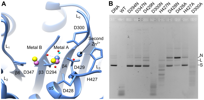
Figure 3.
Metal binding sites. (A) Active site of the G20c nuclease complex with Zn2+. The two Mn2+ ions, taken from structure superposition with SPP1 and HCMV nuclease-Mn2+ complexes are in magenta and yellow, respectively. The water nucleophile is shown in cyan. (B) In vitro nuclease assays. Activity is shown for the wild-type and active site mutant proteins. N, nicked; L, linear; S, supercoiled DNA.

