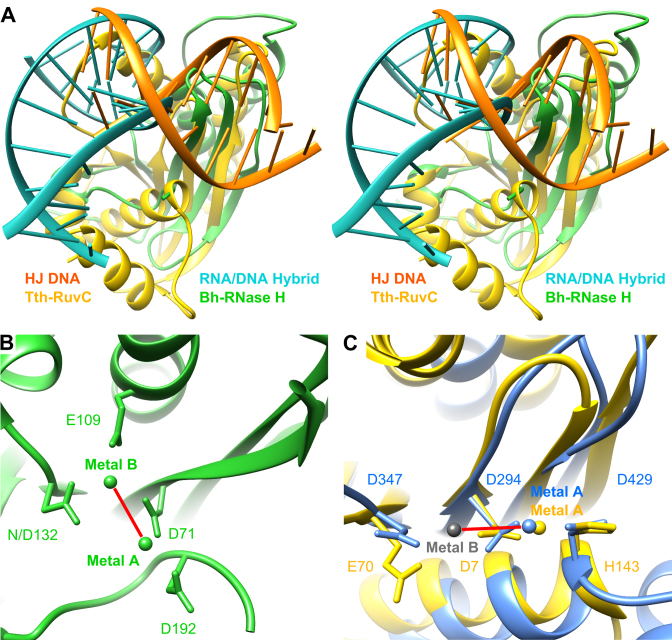
Figure 5.
Comparison of metal location and DNA orientation. (A) Stereo view showing the superposition of nucleic acid complexes of Bacillus halodurans RNase H (green; RNA/DNA hybrid, cyan; PDB accession code 1ZBI) and Thermus thermophilus RuvC (yellow; Holliday junction DNA, orange; 4LD0). (B) Active site of Bh-RNase H nuclease with bound metal ions. (C) Close-up view at the active site in superposed G20c (blue, 5M1N) and Tth-RuvC (yellow, 4EP4) nucleases shown in the same orientation as (B). Bound metal ions (in both structures only at site A) are in corresponding colors. The gray sphere indicates the site B position (as in Lactococcus phage bIL67 RuvC, 4KTZ). Note the different relative orientation of the two metals in (B) and (C), indicated by red line.

