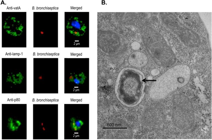
Fig 2. Visualization of B. bronchiseptica within D. discoideum.
(A) Fluorescent confocal imaging of B. bronchiseptica within D. discoideum. D. discoideum were exposed to B. bronchiseptica RB50 pLC018 (mCherry, red) for 1 h at a multiplicity of infection (MOI) of 100 prior to 1 h gentamicin treatment. D. discoideum were then fixed in 4% paraformaldehyde and permeabilized with 0.1% saponin prior to incubation with the following primary antibodies: mouse monoclonal anti-vatA, rat monoclonal anti-lamp-1, and mouse monoclonal anti-p80. Secondary antibodies, specifically goat anti-mouse and goat anti-rat, were conjugated to either fluorescein or a green fluorescent dye (green). D. discoideum were also stained with DAPI for visualization of nuclei (blue). Z-stacks were captured at 60× magnification and zoomed to 300%. Individual panels and merged fluorescent images are displayed. (B) Electron microscopy images of intracellular B. bronchiseptica within D. discoideum. D. discoideum were exposed to B. bronchiseptica RB50 for 1 h at a MOI of 100 prior to 1 h gentamicin treatment. Following exposure and gentamicin treatment, D. discoideum harboring intracellular bacteria were fixed for 1 h in 2% glutaraldehyde and then processed for transmission electron microscopy. The image shows a single intracellular bacterium (arrow) within D. discoideum.

