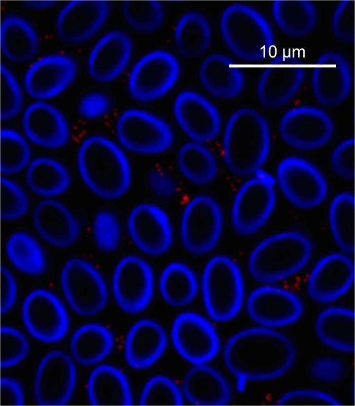
Fig 5. B. bronchiseptica localizes to the amoeba sorus, distributed between the D. discoideum spores.
Confocal microscopy image of D. discoideum sori grown on a lawn of B. bronchiseptica RB50 pLC003 (mCherry, red) at 60× magnification and zoomed to 200%. Amoeba spores were stained with calcofluor (blue).

