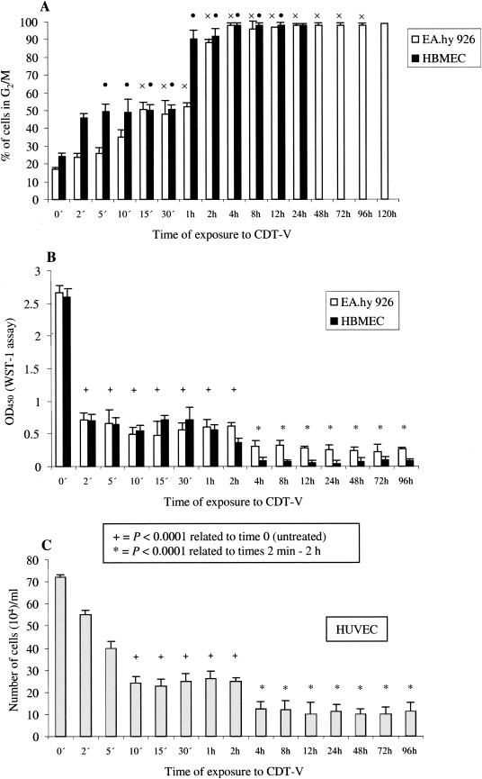
FIG. 6.
Minimum length of exposure to CDT-V required for G2/M arrest and inhibition of proliferation in endothelial cells. Cells were exposed to CDT-V (8 CD50/ml) for the indicated times, the toxin then was removed, and cells were washed and incubated in medium without CDT-V. G2/M arrest (A) was analyzed by flow cytometry 24 h (HBMEC) or 120 h (EA.hy 926 cells) after the addition of CDT-V. The percentages of the total numbers of cells arrested in G2/M after 24 h (HBMEC) or 120 h (EA.hy 926 cells) are shown, and the exposure times which resulted in G2/M arrest in significant proportions (≥50%) of EA.hy 926 cells (×) and HBMEC (•) also are shown. Cell proliferation was measured by the WST-1 assay (EA.hy 926 cells and HBMEC) (B) or trypan blue exclusion (HUVEC) (C) 96 h after the addition of the toxin. Cells at 0 min were not exposed to CDT-V and were cultured in medium only. Differences in mean optical densities at 450 nm (OD450) (EA.hy 926 cells and HBMEC) and cell numbers (HUVEC) were compared with the paired t test, and the P values are shown. All data are means and standard deviations from three independent experiments.

