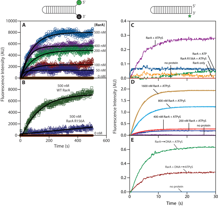
Figure 7.
RarA separates the strands of a double-stranded DNA duplex. (A) The fluorescence intensity of a 6-FAM/3-Dabcyl labeled DNA duplex was measured for 500 s after the addition of RarA in the presence of ATP. (B) The fluorescence intensity of the same DNA substrate was measured over the course of 500 s after addition of either wild-type RarA or RarA R156A variant (0.5 μM) in the presence of ATP. (C) RarA (0.4 μM) was pre-incubated with a 2-aminopurine labelled DNA substrate (0.1 μM molecules; 4.6 μM nucleotides). Fluorescence intensity of 2-aminopurine was measured for 30 seconds following addition of indicated nucleotide cofactors. (D) Varying concentration of RarA were pre-incubated with a 2-aminopurine labelled DNA substrate (0.1 μM molceules; 4.6 μM nucleotides). Fluorescence intensity of 2-aminopurine was measured for 30 seconds following addition of ATPγS. (E) RarA (0.4 μM) was added to a solution containing 2-aminopurine DNA substrate (0.1 μM molceules; 4.6 μM nucleotides) and ATPγS. In a previous experiment, ATPγS was added to a solution containing RarA (0.4 μM) and 2-aminopurine DNA substrate (0.1 μM molceules; 4.6 μM nucleotides). Fluorescence intensity of 2-aminopurine was measured over the course of 30 s in both experiments. All values represent the average of at least three replicate experiments.

