Figure 4.
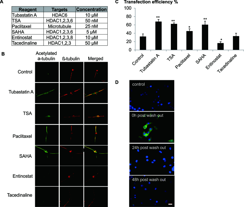
Effect of HDAC6 inhibition on the microtubule stabilization is specific and temporal. (A) Cells were treated with HDACi and Palitaxel at the indicated concentration. (B) Differentiated Neuro2A cells (RA) were exposed to various HDAC inhibitors and Paclitaxel for 2 h. Cells were then fixed with 4% formaldehyde and co-stained for acetylated α-tubulin (Green) and β-tubulin (Red). Confocal images were shown (100× magnification). (C) Differentiated Neuro2A cells were transfected in the presence of DOPE/CHEMS and various microtubule stabilizers for 12 h. Cell treated with only DOPE/CHEMS served as the control. Transfection efficiency was quantified by FACS analysis 48 h later and presented as mean ± SD (n = 3). Significant differences in transfection efficiencies between control and HDACi/Palitaxel treated cultures were calculated using the two tailed Student's t-test. *P < 0.05; **P < 0.005. (D) Neuro2A cells were treated with Tubastatin A for 12 h (0 h washout) and the media was then replaced with complete media. At various time points, cells were fixed with 4% formaldehyde and stained for acetylated α-tubulin (green) and nucleus (Hoechst stain, blue). Cells not treated with Tubastatin A were used as a control. Representative images were shown. Bar represents 20 μm.

