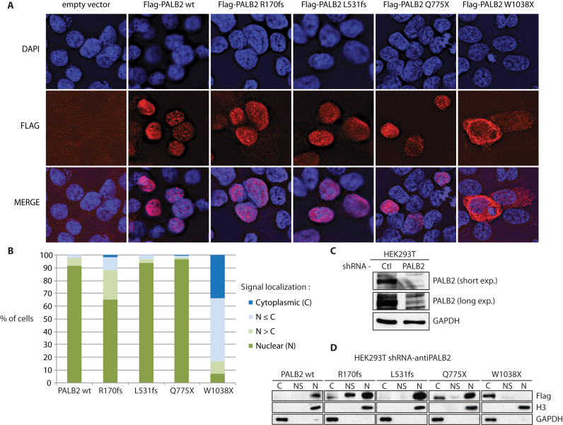
Figure 3.
Cellular localization of PALB2 mutant proteins. (A) Immunofluorescence of mutant forms of Flag-tagged PALB2. DAPI (blue), anti-Flag (red), and the merge picture are shown. All mutants except W1038X were mainly nuclear. (B) Quantification of the cytoplasmic and nuclear accumulation of PALB2 mutants. Experiments were performed in quadruplicate. (C) Knockdown of endogenous PALB2 in HEK293T cells by expressing constitutively a shRNA against PALB2. (D) Analysis of the cellular localization of PALB2 mutant forms by cellular fractionation. The cytoplasmic (C), nuclear soluble (NS) and chromatin (N) fractions are shown. Blots against the GAPDH and histone H3 proteins are shown as controls.

