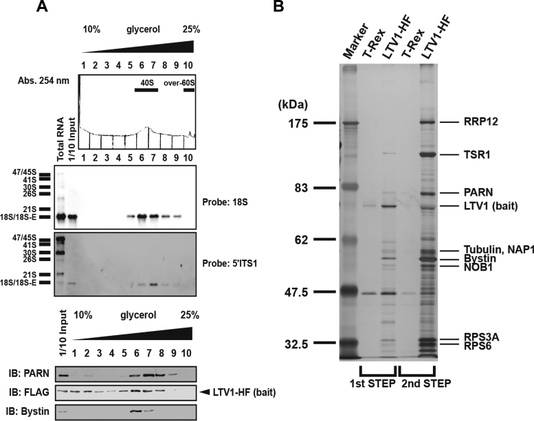
Figure 1.
Purification of human LTV1-HF-associated pre-40S particles using two-step affinity purification. (A) The LTV1-associated complex prepared by two-step purification was separated into 10 fractions by glycerol gradient centrifugation (10–25% glycerol, top). The eluate was monitored at 254 nm. RNAs in each fraction were detected by northern blotting with a probe for 18S or 5΄ITS1. RNA sizes are indicated to the left. Proteins eluted in each fraction were detected by immunoblotting (IB) with an antibody against PARN, FLAG, or Bystin. (B) LTV1-associated proteins isolated from T-Rex or T-Rex cells expressing LTV1 (LTV1-HF) were separated by SDS-PAGE and visualized by silver staining. Proteins were identified by LC–MS/MS after in-gel trypsinization. Molecular mass markers (kDa) are indicated to the left, and the identified protein names are shown on the right. Lane 1, marker proteins; lane 2, control for the first step of purification; lane 3, proteins isolated by the first step of purification (10%); lane 4, control for the second step of purification and lane 5, proteins isolated by the second step of purification (100%).

