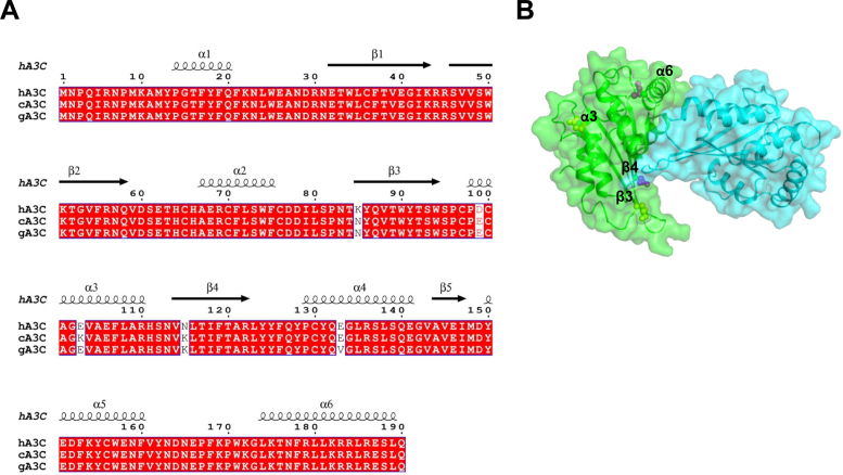
Figure 2.
Sequence alignment and structural analysis of A3C. (A) Sequence alignment of hA3C, cA3C and gA3C with amino acid differences shown in white. The sequence alignment was performed by a Clustal Omega multiple sequence alignment (75) and plotted using the program ESPript (76). (B) Surface representation of a hA3C dimer from the crystal structure (PDB: 3VOW). Amino acids unique to hA3C that are potentially involved in the dimer interface are shown in purple (α-helix 6, S188; β-strand 4, N115) and other amino acids unique to hA3C are shown in yellow (β-strand 3, K85; α-helix 3, D99).

