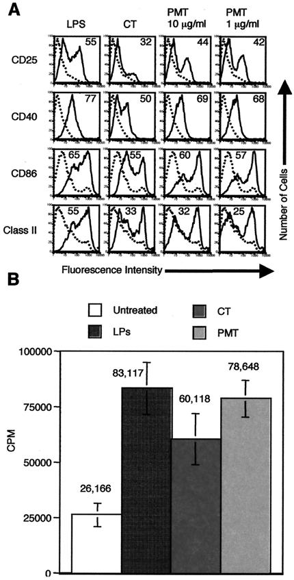
FIG. 4.
PMT activates murine BMDC to mature and increases their ability to present alloantigen to T cells in the allogeneic T-cell response. A. Cell surface expression of the indicated markers on untreated BMDC (dotted histograms) or BMDC treated with the indicated agonist (solid histograms). Day 6 BMDC were incubated with 1 μg of LPS or CT/ml or PMT at 1 or 10 μg/ml for 24 h. The cells were harvested and stained for single-color flow cytometry with PE-anti-CD25, PE-anti-CD40, PE-anti-CD86, or PE-anti-14-4-4S (class II MHC). Data are representative of one experiment of three performed on cells from different mice with similar results. B. Total C57BL/6 T cells were plated at 105 cells/well in 96-well U-bottom plates in T-cell medium. Day 7 BMDC were left untreated or were activated by a prior 24-h incubation with 1 μg of LPS, CT, or PMT/ml. Untreated and activated BALB/c BMDC were washed, and then 1,000 cells were added to the C57BL/6 T cells. Experiments for each condition were performed in triplicate. Proliferation was determined at day 5 by pulsing the cells with 1 μCi of [3H]thymidine per well for the last 18 h of culture. Thymidine incorporation was measured with a Packard Matrix 96 direct beta counter. Data are means and standard deviations for mixtures of BMDC and T cells generated from three separate mice. CPM, counts per minute.

