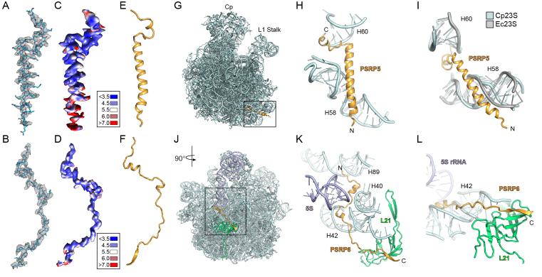
Figure 5.
Localization of PSRP5 and PSRP6 on the chlororibosome. (A and B) Cryo-EM electron density (mesh) with molecular models for (A) PSRP5 and (B) PSRP6. (C and D) Cryo-EM electron density for (C) PSRP5 and (D) PSRP6 colored according to local resolution. (E and F) Molecular models showing secondary structure for (E) PSRP5 and (F) PSRP6. (G) Binding site of PSRP5 (gold) on the LSU (cyan). (H and I) Interaction between PSRP5 (gold) and H58 and H60 of the cp23S rRNA (cyan), with (I) comparison of different conformation of H58 from the Ec23S rRNA (gray). (J) Binding site of PSRP6 (gold) on the LSU (cyan). (K and L) Interaction between the N-terminus of PSRP6 (gold) and H40, H42 and H89 of the cp23S rRNA (cyan), and (L) the C-terminus of PSRP6 (gold) with the cpL21 (green).

