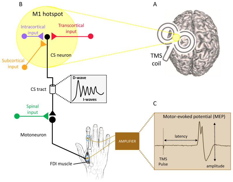
Figure 1. Transcranial magnetic stimulation (TMS) as a probe of corticospinal excitability.
A. The TMS coil is placed over primary motor cortex (M1) at the “hotspot” (depicted in yellow), the position at which the largest motor-evoked potentials (MEPs) can be recorded in the EMG signal from a targeted muscle. B. TMS over M1 activates corticospinal (CS) neurons directly or indirectly via the stimulation of intracortical circuits that project to CS neurons. Transcortical inputs from premotor, prefrontal and parietal cortices, as well as axons of subcortical cells projecting onto M1 are also activated by TMS over M1. Depending on the position and intensity of stimulation, a series of descending volleys (D- wave and I-wave) are transmitted from M1 to the motorneurons in the spinal cord. These signals are further influenced by inputs at the spinal level before they jointly give rise to an MEP in the targeted, contralateral muscle (first dorsal interosseus [FDI] in the present example). C. The MEP is a bi-phasic response recorded from a targeted muscle via electrodes placed on the surface of the skin. It has a latency of approximately 18 ms after the TMS pulse when elicited in hand muscles. While the latency is relatively invariant, the peak-to-peak amplitude fluctuates, reflecting the sum of cortical, subcortical, and spinal contributions to the descending signals to the muscle.

