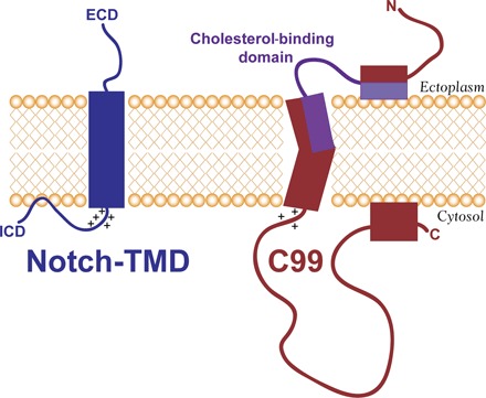
Fig. 7. Summary of differences between C99 and the Notch-TMD.

ECD and ICD are the large Notch extracellular and intracellular domains, respectively (see Fig. 1). The general locations of the key residues in the cholesterol binding site of C99 are indicated in purple. Not illustrated here is the propensity of C99, but not the Notch-TMD, to dimerize. Also not illustrated here is the fact that, if the membrane shown in this figure were thinned, the Notch-TMD is predicted by the results of this work to adjust by tilting with respect to the bilayer normal, whereas C99 would remain untilted, with the N-terminal end of its TMD jutting out into the ectoplasm.
