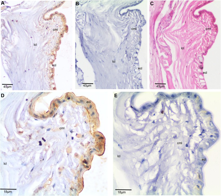
Fig 3. Expression of E. eugeniae TCTP in intact posterior segments of E. eugeniae.
(A) Immunohistochemistry with anti-TCTP antibody using the intact posterior segments of E. eugeniae. It shows mere expression of TCTP at the epithelial cell region (B) The pre-immune serum treated sections used as a control, it didn’t show the signals. (C) The consecutive section of IHC sample was stained with eiosin and haemotoxylin. (D) 40X Magnified image of panel A shows the expression of TCTP protein ECL layer. (E) 40X magnified image of Panel B, show no positive signals.

