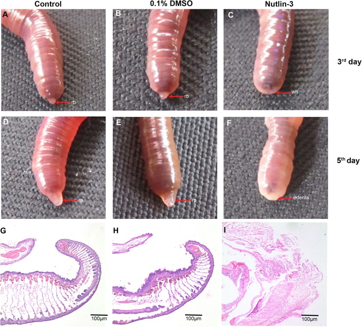
Fig 6. Regeneration of nutil-3 injected worm.
(A) Non injected control animals, it formed the regeneration blastema on the 3rd day. (B) 0.1% of DMSO injected worm, it formed the regeneration blastema on the 3rd day (C) The worm injected with 5 mg/Kg concentration of nutlin-3 was amputated and maintain for regeneration. The worm on 3rd day shows no regeneration blastema. (D) Control 0.1% of DMSO injected worm formed the regeneration blastema. (E) Non injected control animal formed the blastema as expected. (F) The nutlin-3 injected worm on 5th day fails to form blastema even in fifth day. (G) Histological image (4X) of non injected worm tissue, it shows well developed regenerated part. (H) Histological image (4X) of 5th day control 0.1% DMSO injected worm tissue, it shows well developed regenerated part. (I) Histological image (4X) of 5th day nutlin-3 treated worm tissue, the cells are loosely packed at the amputated region.

