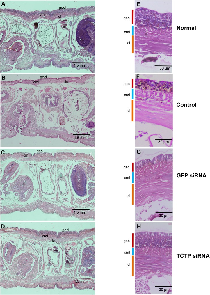
Fig 11. Cellular abnormality of clitellum during TCTP RNAi.
(A and E) 3X and 10X magnified histological image of intact clitellum shows thick glandular epithelial cell layer (gecl) (B and F) 3X and 10X magnified histological image of the 5th day regenerating worm, it shows drastic reduction of glandular epithelial cell layer in the clitellar segments. (C and G) 3X and 10X magnified histological image of the 5th day regenerating GFP siRNA injected worm, it shows drastic reduction of glandular epithelial cell layer in the clitellar segments. (D and H) 3X and 10X magnified histological image of the 5th day TCTP siRNA injected worm, it retains the structure of control worm’s clitellar glandular epithelial cell layer.

